Key Takeaways:
- Uneven shoe wear, recurring one-sided pain, or balance issues signal that a professional gait check can prevent bigger injuries.
- Kinotek 3-D motion capture, pressure mapping, and EMG pinpoint exactly where and when each joint or muscle misfires.
- Regular warm-ups, year-round strength work, timely shoe changes, and periodic re-tests keep your gait efficient and pain-free.
Every footstep follows a pattern. When that pattern shifts, even a little, your body tries to adjust. A sore knee, a tight calf, or an aching back can be the first signs that something in your gait needs attention. Gait is the way you walk or run. When it works well, you use less energy and avoid extra stress on joints. When it falters, pain and injury often follow.
Understanding Gait Basics
A normal gait cycle has two broad phases:
Stance: the foot is on the ground and carries weight.
Swing: the foot lifts and swings forward.
Each phase includes smaller moments, heel strike, mid‑stance, toe‑off. Muscles fire in sequence; joints flex and extend. A well‑timed cycle saves energy. A flawed one wastes it and puts strain where it doesn’t belong. Gait analysis measures those flaws so they can be fixed. Researchers define gait analysis as a systematic study of human motion using observation and instruments to track joint angles, forces, and muscle activity.
Common Gait Faults and Why They Happen
Understanding common gait abnormalities is crucial for diagnosing underlying musculoskeletal issues and developing effective treatment plans. These deviations from a typical walking pattern often indicate specific muscle weaknesses, structural imbalances, or pain points.
Over-pronation
Characterized by the arch of the foot collapsing inward excessively during the stance phase of gait. This often presents as a flattening of the medial longitudinal arch, where the ankle rolls inward.
Likely Causes:
- Weak Tibialis Posterior Muscle: This muscle is a primary dynamic stabilizer of the arch. When weak, it struggles to maintain the integrity of the arch.
- Low Arches (Pes Planus): Individuals with inherently low or flat arches are more predisposed to over-pronation.
- Worn Shoes: Footwear that lacks adequate arch support or has an excessively worn-down medial sole can exacerbate or cause over-pronation.
Consequences:
Can lead to a cascade of issues including shin splints, patellofemoral pain syndrome, plantar fasciitis, and even lower back pain due to altered biomechanics up the kinetic chain.
Under-pronation (Supination)
In contrast to over-pronation, the foot stays predominantly on its outer edge during the stance phase, with insufficient inward rolling. The arch remains high and rigid.
Likely Causes:
- High, Rigid Arches (Pes Cavus): Individuals with naturally high and inflexible arches are more prone to supination. These arches do not absorb shock effectively.
- Stiff Ankles: Limited ankle dorsiflexion or general ankle rigidity can prevent the foot from adequately pronating, forcing it to remain on its lateral border.
Consequences:
Often results in poor shock absorption, leading to stress fractures (especially in the fibula or metatarsals), ankle sprains (due to instability on the outer edge of the foot), iliotibial band syndrome, and knee pain.
Trendelenburg Gait
Identified by the dropping of the hip on the opposite side of the stance leg during the single-leg support phase of gait. For example, if standing on the right leg, the left hip will drop.
Likely Causes:
- Weak Gluteus Medius Muscle: The gluteus medius is a key hip abductor responsible for stabilizing the pelvis during walking. Weakness leads to the unsupported side of the pelvis dropping.
- Pelvic Instability: General instability in the sacroiliac joint or lumbar spine can also contribute.
Consequences:
Can lead to compensatory trunk leaning (leaning towards the affected side) to maintain balance, lower back pain, knee pain, and further gait deviations.
Antalgic Gait
A characteristic gait where the individual significantly shortens the stance phase on the painful leg, quickly shifting weight to the unaffected leg to minimize discomfort.
Likely Causes:
- Foot Pain: Conditions like plantar fasciitis, bunions, metatarsalgia, or fractures.
- Knee Pain: Arthritis, meniscal tears, ligamentous injuries, or patellofemoral pain.
- Hip Pain: Osteoarthritis, bursitis, labral tears, or hip impingement.
Consequences:
While providing immediate pain relief, it alters the normal biomechanics, leading to increased stress on the unaffected limb and potential secondary pain or injury in other joints over time.
Toe-in Gait (In-toeing)
The feet point inward during walking, often described as “pigeon-toed.”
Likely Causes:
- Excessive Hip Internal Rotation: This is common in children and can be due to femoral anteversion (a twist in the femur).
- Muscle Tightness: Tight hip internal rotators or overactive adductor muscles can contribute.
Consequences:
Can affect balance and coordination, lead to frequent tripping, and potentially contribute to knee and hip problems if persistent into adulthood.
Toe-out Gait (Out-toeing)
The feet point outward during walking, sometimes significantly.
Likely Causes:
- Tight External Rotators of the Hip: Muscles like the piriformis or obturators can become tight, externally rotating the femur.
- Compensation for Hip Stiffness: If hip flexion or internal rotation is limited (e.g., due to hip arthritis), the body may compensate by externally rotating the hip to achieve necessary clearance or range of motion during gait.
- Tibial Torsion: A twist in the tibia (shin bone) can also cause out-toeing.
Can alter weight distribution across the foot, potentially leading to bunions or hammertoes, and can place abnormal stress on the knees and hips.
Vaulting Gait
Occurs when an individual rises up on the toes of their support leg (the unaffected leg) to provide extra clearance for the swing leg to pass through without dragging.
Likely Causes:
- Leg-Length Difference: If one leg is significantly shorter than the other, the body will “vault” on the longer leg to clear the shorter one during its swing phase.
- Limited Knee Flexion: If the knee of the swing leg cannot bend sufficiently (e.g., due to a knee brace, stiffness, or pain), the individual will vault on the stance leg to elevate the pelvis and clear the straight leg.
Consequences:
Can lead to increased energy expenditure during walking, uneven weight distribution, and potential strain on the ankle and foot of the vaulting leg.
Weakness, stiffness, or poor technique usually sit at the root. Treating only the pain ignores that root and invites the problem back.
Who Should Consider a Gait Assessment
Who benefits from a gait check? Anyone who spends serious time on their feet, distance runners, athletes who cut and jump, healthcare or retail workers, and older adults eager to stay steady, can learn from it. It’s equally helpful if you’re recovering from lower‑limb surgery or dealing with foot, knee, hip, or back pain that keeps cycling back. In short, if movement is central to your day and discomfort tags along, a gait assessment belongs on your to‑do list.
Clues Your Gait Needs Attention
Pain is just one messenger, and it often speaks late. Earlier warnings include uneven wear on your shoes, blisters that pop up in strange places, or a limp a friend spots before you feel it. Repeating injuries on the same side of the body or a nagging sense you’re working harder than usual to stay balanced are other red flags. Treat these hints as an invitation to get checked before the issue settles in.
Why Early Analysis Helps
Fixing a gait issue is like realigning a car wheel. Quick action saves the tires. Small corrections, new shoes, targeted exercises, or different pacing, stop pain before it locks in. Waiting can add scar tissue, reduce mobility, and shift stress to other joints.
Tools and Technology
Modern labs use a blend of devices:
- High‑speed cameras track joint angles in slow motion.
- Pressure plates show how force spreads under each foot.
- Marker‑based 3D capture maps full‑body movement.
- In‑shoe sensors record real‑world strides.
- Surface EMG reads muscle timing.
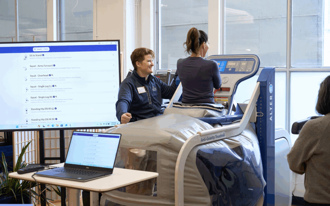
At Avid Sports Medicine, sessions run on Kinotek Movement Software, which captures 3D joint data and muscle engagement in real time
Gait Analysis Process
- Interview – Medical history, training load, pain points, and goals.
- Baseline Exam – Range of motion, strength, flexibility.
- Motion Capture – Walk or run on an instrumented treadmill. Cameras and Kinotek sensors log each move.
- Pressure Mapping – Plates or in‑shoe sensors reveal force patterns.
- Data Review – A clinician breaks down findings frame by frame.
- Action Plan – Tailored exercises, technique cues, and footwear advice.
- Follow‑up – Re‑test after four to six weeks to confirm progress.
Gait Analysis Process
Interview: Medical history, training load, pain points, and goals.
- Baseline Exam: Range of motion, strength, flexibility.
- Motion Capture: Walk or run on an instrumented treadmill. Cameras and Kinotek sensors log each move.
- Pressure Mapping: Plates or in‑shoe sensors reveal force patterns.
- Data Review : A clinician breaks down findings frame by frame.
- Action Plan: Tailored exercises, technique cues, and footwear advice.
- Follow‑up: Re‑test after four to six weeks to confirm progress.
Rehabilitation Phases After a Fault Is Found
Phase 1: Protection
Rest painful tissues. Light mobility keeps blood flowing. Ice and gentle compression calm inflammation.
Phase 2: Strength and Control
Strength work now targets the gaps revealed by your assessment. Single‑leg bridges, side‑plank hip lifts, and slow calf raises make up the core of most programs and run two or three times a week. The goal is to firm up control around the hips and ankles so each foot lands under a steady frame.
Phase 3: Re‑integration Re‑integration
Add form drills, high knees, A‑skips, butt kicks, and low‑volume walk‑run intervals. Build tolerance with gradual load.
Phase 4: Performance
Return to hills, speed work, or sport‑specific drills. Deload every fourth week. Keep strength work once a week.
Strategies to Keep a Healthy Gait
Warm‑up and Technique Habits
Start every outing with a brisk five‑minute walk and leg swings to wake tissues and raise body temperature. Pay attention to cadence; nudging your step count a little higher often shortens an over‑stride and softens ground impact.
Ongoing Training Habits
Keep two brief strength sessions in your week so glutes, calves, and core stay reliable. Vary terrain, grass, track, and road, to spread load across tissues, and replace shoes before their midsoles pack down (usually 300–500 miles). Round it out with seven to nine hours of sleep so soft tissue repairs on schedule.
Consistency, not novelty, keeps gait on track.
Your gait is your movement fingerprint. When alignment slips, your body sounds the alarm with discomfort and inefficiency. Gait analysis listens to that signal. It blends advanced tools, like Kinotek, and expert eyes to reveal what each joint, muscle, and footfall is doing. From there, targeted strength, mobility, and technique cues guide lasting change. Prevention is a steady commitment to good habits, warm‑ups, strength work, and smart training loads. If something feels off, trust that instinct. Early testing is easier than long rehab.
Book Your Gait Analysis San Francisco Session at Avid Today
Ready for data‑driven answers to pain or performance questions? Schedule a gait analysis with our team today: Avid Sports Medicine. We’ll study your stride, explain the findings, and build a plan that keeps you moving well.